Ultrasound therapy is a non-invasive treatment that uses high-frequency sound waves to support the healing of injured soft tissues, including muscles, tendons, and ligaments. These sound waves operate at frequencies above human hearing and create gentle mechanical vibrations within the body.
In clinical practice, therapeutic ultrasound typically uses frequencies between 1 and 3 megahertz (MHz). As the sound waves move through body tissues, they cause microscopic vibrations at a cellular level. This process can enhance blood circulation, reduce pain and stiffness, and promote the body’s natural healing response.
Although ultrasound therapy can generate mild warmth in the tissues, its primary therapeutic benefits come from its non-thermal effects, which help improve tissue mobility and accelerate recovery.
How Ultrasound Therapy Works
The ultrasound treatment head contains a small crystal attached to a metal plate. When electrical energy is delivered from the ultrasound machine, the crystal vibrates and produces sound waves. These vibrations are delivered at either 1 MHz or 3 MHz, depending on how deep the target tissue is located. Modern ultrasound heads often use harmonic technology, allowing a single applicator to produce both frequencies.
As ultrasound waves pass through the body, they stimulate tissue through processes known as acoustic streaming and stable cavitation. These effects increase cellular activity and enhance the efficiency of tissue repair. Rather than directly healing the tissue, ultrasound acts as a catalyst that encourages the body’s own repair mechanisms.
Effects of Ultrasound Therapy During the Healing Process
Inflammatory Phase
In the early stage of healing, ultrasound therapy increases the activity of key inflammatory cells, including mast cells, platelets, white blood cells, and macrophages. This supports the controlled inflammatory response required to initiate tissue repair.
Proliferation Phase
During the tissue repair and scar formation stage, ultrasound therapy stimulates fibroblasts and blood vessel cells. This encourages the production of new tissue and improves circulation to the injured area, supporting faster and more effective healing.
Remodelling Phase
In the final phase of healing, ultrasound therapy helps improve the quality and structure of scar tissue. It supports proper collagen alignment and promotes the conversion of weaker Type III collagen into stronger Type I collagen, enhancing tissue strength, flexibility, and durability.
Key Benefits of Ultrasound Therapy
Stimulates cellular activity and tissue repair
Supports healing throughout all stages of recovery
Improves scar tissue quality and flexibility
Helps restore normal movement and tissue function
Where and How Ultrasound Therapy Works Best
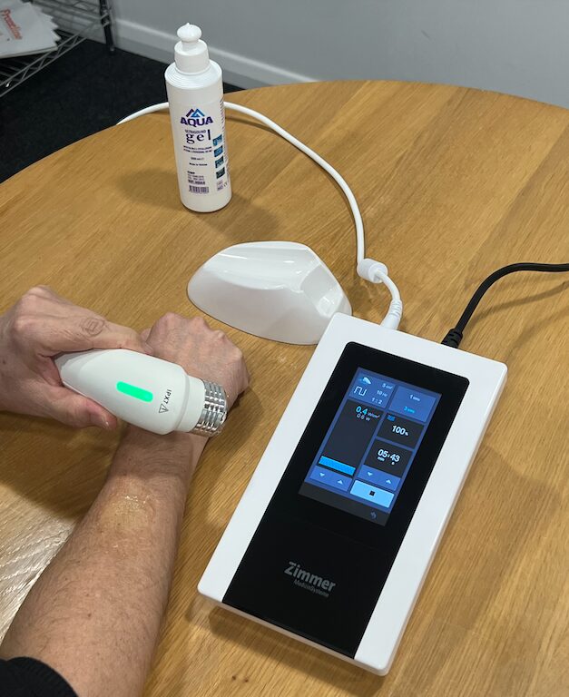
Ultrasound waves cannot travel through air, so a coupling medium is required to transfer the sound waves from the treatment head into the skin. For this reason, a water-based ultrasound gel is applied during treatment.
Water-based gel allows ultrasound energy to pass efficiently into the tissues. Oils and lotions, by contrast, are poor conductors of sound waves and can significantly reduce treatment effectiveness.
Most modern ultrasound machines include contact control technology, which ensures the treatment head maintains proper contact with the skin and gel. This feature helps protect the equipment while ensuring safe and consistent delivery of ultrasound energy.
How Ultrasound Energy Is Absorbed in the Body
Ultrasound energy is absorbed by body tissues in an exponential pattern. This means that tissues closer to the skin absorb more energy, while deeper tissues receive progressively less. For ultrasound therapy to be effective, the energy must reach and be absorbed by the targeted tissue.
To achieve this, treatment parameters such as frequency, intensity, and dosage are carefully selected. These settings ensure the ultrasound penetrates to the appropriate depth and produces the intended therapeutic effect.
Contraindications for Ultrasound Therapy
Ultrasound therapy is generally safe when applied correctly, but there are specific situations where it should not be used or must be used with caution to avoid potential harm.
Absolute Contraindications
Ultrasound therapy must not be applied in the following cases:
Pregnancy (over the abdomen, pelvis, or lower back)
Ultrasound should not be used over the trunk during pregnancy due to potential risks to the developing fetus.
Malignant tumours (cancerous tissue)
Ultrasound may increase blood flow and cellular activity, which could potentially stimulate tumour growth or spread.
Active infection or sepsis
Increased circulation from ultrasound may worsen the spread of infection.
Areas with active bleeding or haemorrhage
Ultrasound can increase blood flow and may exacerbate bleeding.
Thrombophlebitis or suspected deep vein thrombosis (DVT)
Ultrasound may increase the risk of dislodging a clot.
Over the eyes
The eye is highly sensitive to ultrasound energy and can be damaged.
Over the brain or spinal cord (post-laminectomy)
Especially when protective bone structures are absent.
Over a pacemaker or implanted electronic devices
Ultrasound may interfere with device function.
Relative Contraindications / Precautions
Ultrasound may be used only with caution or modified settings in the following situations:
Reduced sensation or impaired circulation
Patients may not feel excessive heat, increasing the risk of tissue damage.
Areas of plastic or metal implants
Modern implants are usually safe, but dosage should be adjusted and monitored.
Epiphyseal plates (growth plates) in children
Avoid direct application over active growth plates.
Severe osteoporosis
Fragile bones may be more susceptible to injury.
Over reproductive organs
Avoid direct application unless clinically justified and appropriately dosed.
Scar tissue with reduced sensitivity
Requires careful monitoring to avoid overheating.
Additional Safety Considerations
Ultrasound should not be applied over air-filled structures, such as the lungs or bowel.
Continuous ultrasound should be used cautiously to prevent excessive thermal effects.
Treatment parameters (frequency, intensity, duty cycle) should always be selected based on tissue depth and clinical presentation.