Anatomical Models
Anatomical models for physiotherapy, sports therapy, chiropractic, and osteopathy practices. The range covers life-size skeletons, spine and vertebral column models, skull models, and peripheral joint models, including shoulder, hip, knee, elbow, hand, wrist, and foot. Most models are fully articulated and come on a stand, making them practical for both desktop reference and patient-facing consultation use. If you need anatomical charts alongside your models, see our human anatomical charts range.
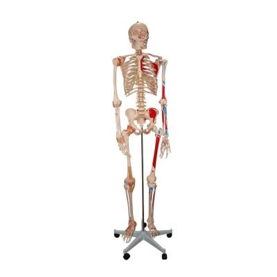
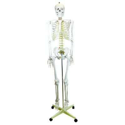
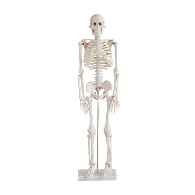
Explore our extensive collection of anatomical models, designed to enhance medical education and help students master human anatomy. Many of our models are life-size and fully articulate, providing a hands-on, realistic understanding of each body part.
These three-dimensional, tactile models are perfect for students, universities, colleges, GPs, and clinics, offering an engaging way to visualize and study anatomy.

Why a physical model still earns its place in any clinic
You can describe a disc herniation in detail. You can draw it. You can pull up an image on a screen. But nothing closes a consultation faster than putting a lumbar spine model in a patient's hands and showing them exactly what's happened and why it hurts. A randomised controlled trial found that patients shown an anatomical model during a consultation reported significantly higher satisfaction than those given a verbal explanation alone, and rated their clinician's interpersonal skills more highly too. The model does part of the work for you.
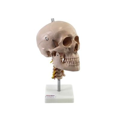
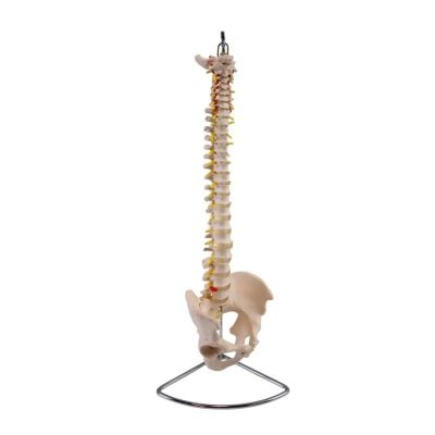
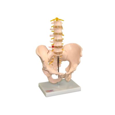
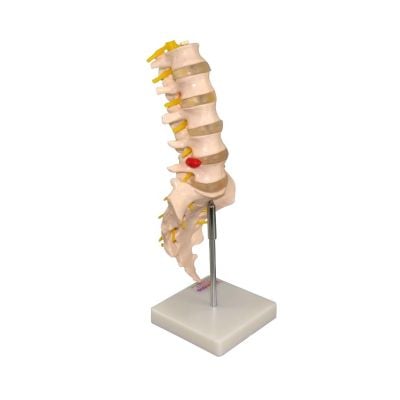
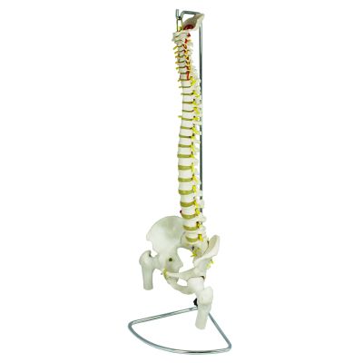
Spine models
Your patients don't read MRI reports. But they do understand when you put a lumbar model in front of them, point to the disc, and show them exactly what's being compressed and why bending forward makes it worse. A flexible vertebral column lets you demonstrate load, movement, and the logic of your treatment approach in under a minute. If disc pathology is a regular part of your caseload, get the prolapsed disc version.
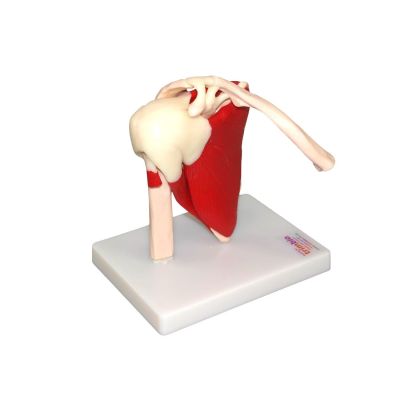
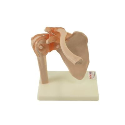
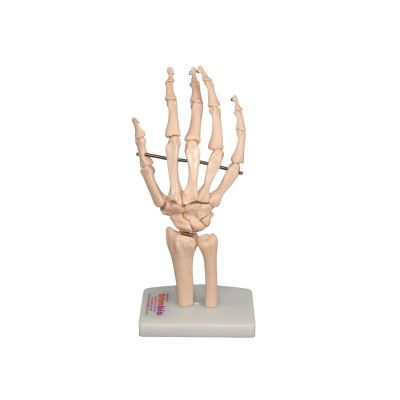
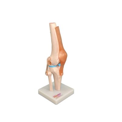
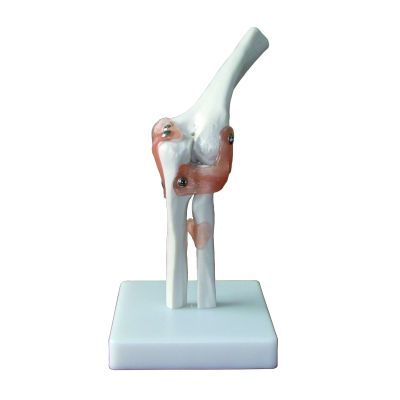
Joint models
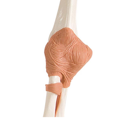
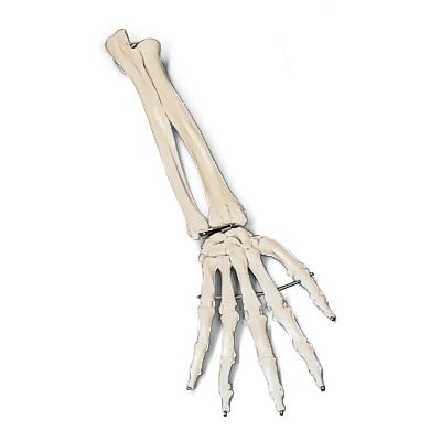
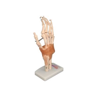
The functional models here move, which is the point. The knee model lets you take a patient through ligament behaviour across a full range, useful for ACL, meniscal, and valgus loading conversations that otherwise take twice as long to explain verbally. The shoulder model has the rotator cuff intact, so impingement and instability presentations are straightforward to show rather than describe. If you treat a lot of hands and wrists, the lifesize model with ligaments covers TFCC and carpal mechanics well enough to support a proper clinical explanation rather than a rough sketch on the couch roll.
Browse our anatomical charts, electrotherapy equipment, treatment couches, and consumables.
FAQs
Is a functional knee model accurate enough to explain meniscal pathology?
Yes. The functional knee model shows ligament behaviour and joint mechanics through a full range of motion. For meniscal presentations, it gives you enough structural context to show the patient where the involvement is and why certain movements load the area. It won't replace an MRI conversation, but it makes that conversation shorter.
Can I use a lumbar spine model to explain the difference between a prolapse and a bulge?
Yes, and it's one of the clearest ways to do it. The prolapsed disc version shows the degree of nuclear displacement in a way that patients immediately grasp. Most clinicians find that it reduces the number of follow-up questions significantly.
My patients often confuse referred pain with the actual injury site. Does a spine model help?
Considerably. Showing a patient where the nerve root exits and tracing the referral pattern on a model is far more effective than describing dermatomes verbally. The models with removable vertebrae let you isolate the segment you're discussing.
Is the shoulder model detailed enough to explain rotator cuff impingement vs a full thickness tear?
The shoulder model shows the cuff attachments and sub-acromial space clearly enough for an impingement explanation. For a full thickness tear conversation, it works well alongside imaging rather than as a standalone.
My patient doesn't understand why their posture is loading their discs differently when sitting. What model helps most?
The flexible vertebral column. You can physically load it into flexion and show what happens to the posterior disc and facet joints in a slumped sitting position versus neutral. Patients who've been told to "sit up straight" for years and never understood why tend to find this useful.
How durable are these anatomical models in a busy clinic?
The hard plastic construction handles regular patient handling well. The painted skeleton and functional joint models are built for clinical environments rather than display cases.